Click on the links below.
Listed below are some of the other features that suggest early pregnancy failure:
- An empty gestation sac: especially after 8 weeks, this means that the pregnancy is unlikely to continue
- The gestation sac may be large but the embryo small, which often heralds imminent demise
- The shape of the gestation sac may be irregular and pointed at one or both ends
- The uterus may be empty – a complete miscarriage
- The gestation sac may contain blood, represented by lots of irregular echoes. Be careful – sometimes there may be bleeding between the membranes (so called subchorionic haemorrhage); although this is not a normal finding, many pregnancies with this feature will continue to the delivery of a normal baby
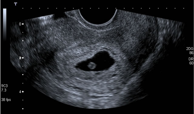
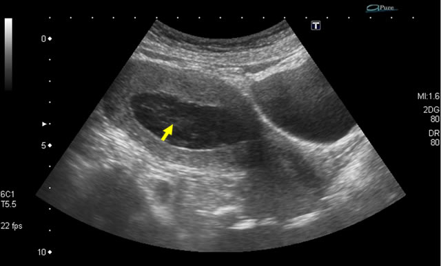
Below is an empty gestation sac with no embryo or yolk sac. Note that there is no obvious trophoblastic layer and that the gestation sac is somewhat flattened and pointed.
Below is an empty gestation sac with no embryo or yolk sac. Note that there is no obvious trophoblastic layer and that the gestation sac is somewhat flattened and pointed.
Below is an empty gestation sac with no embryo or yolk sac. Note that there is no obvious trophoblastic layer and that the gestation sac is somewhat flattened and pointed.

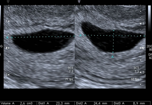
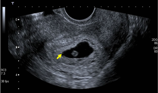
This image shows a normal early gestation. The trophoblastic region surrounding the gestation sac is bright, as expected, and there is a yolk sac. The embryo is not seen but if you did not see this as you scanned through the gestation sac, it would be important to scan again in a week to assess whether the pregnancy had grown and whether you could se a heartbeat, hus confirming that this was a live gestation.
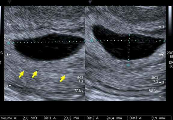
Note that one end of the gestation sac is slightly pointed. This is a sign that is of limited value. It can be seen in normal pregnancies but is more common in a non live pregnancy.
This image shows a normal early gestation. The trophoblastic region surrounding the gestation sac is bright, as expected, and there is a yolk sac. The embryo is not seen but if you did not see this as you scanned through the gestation sac, it would be important to scan again in a week to assess whether the pregnancy had grown and whether you could se a heartbeat, hus confirming that this was a live gestation.
Note that one end of the gestation sac is slightly pointed. This is a sign that is of limited value. It can be seen in normal pregnancies but is more common in a non live pregnancy.
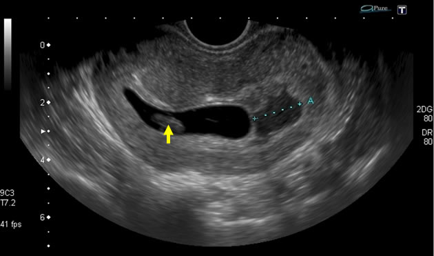
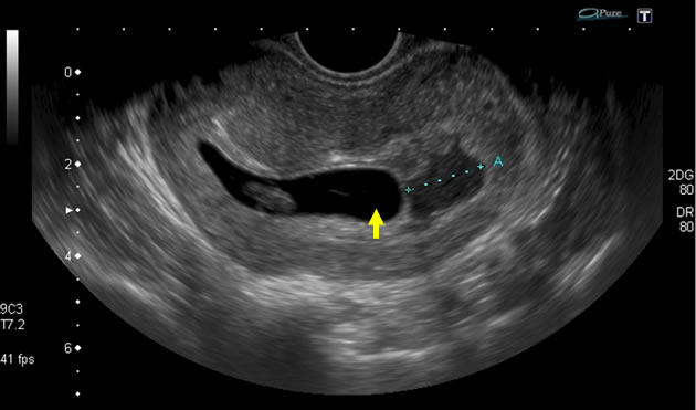
This image shows a gestation sac within the uterus but the wall of the sac is thin and there is no trophoblastic response. There are echoes within the gestation sac, representing blood.
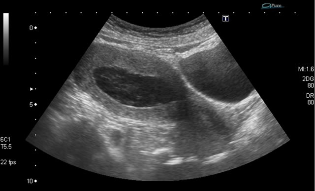
This image shows a gestation sac within the uterus but the wall of the sac is thin and there is no trophoblastic response. There are echoes within the gestation sac, representing blood.
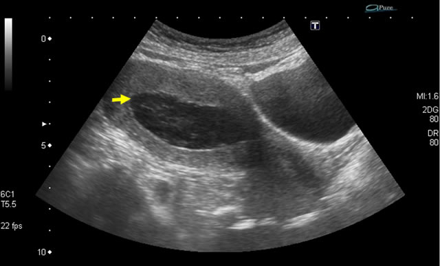
This image shows a gestation sac within the uterus but the wall of the sac is thin and there is no trophoblastic response. There are echoes within the gestation sac, representing blood.
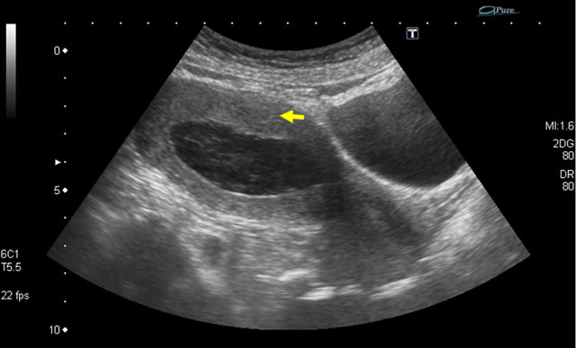
This image shows a gestation sac within the uterus but the wall of the sac is thin and there is no trophoblastic response. There are echoes within the gestation sac, representing blood.
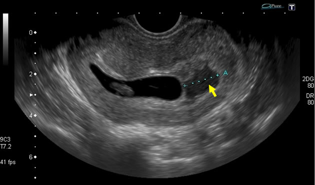
The embryo, gestation sac and chorionic membrane can be clearly seen in this image. However, note the subchorionic haemorrhage.

The embryo, gestation sac and chorionic membrane can be clearly seen in this image. However, note the subchoironic haemorrhage.
The embryo, gestation sac and chorionic membrane can be clearly seen in this image. However, note the subchorionic haemorrhage.
The embryo, gestation sac and chorionic membrane can be clearly seen in this image. However, note the subchorionic haemorrhage.
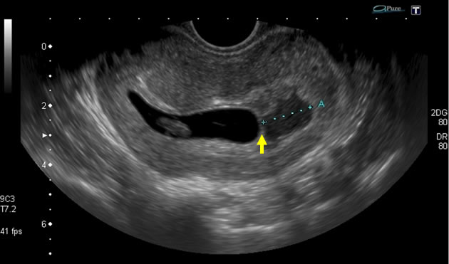
The embryo, gestation sac and chorionic membrane can be clearly seen in this image. However, note the subchorionic haemorrhage.