Technique for Performing BPD Measurement
Click on the links and on the play button to watch the video.
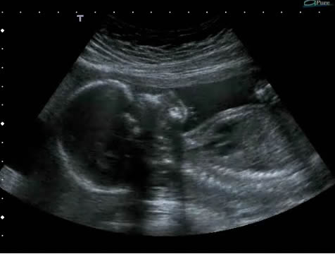
Example 1
In Video 1, note how the baby is first shown in sagittal section, facing towards the ultrasound probe. The sonographer rotates the probe from the sagittal to the axial and then moves the probe so that the scan plane is through the parietal eminences of the fetal skull and the internal anatomy of the fetal brain is shown.
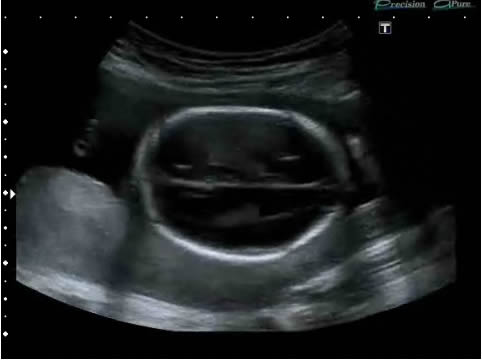
Example 2
In Video 2, note that the section achieved is axial.
On the next page, we shall explore some of the crucial anatomy that will allow you to identify this plane regularly while you are scanning and thus reliably produce the same cross-section for BPD measurement.
Flash video player loading... Flash plugin required.
| Video 1 |
Flash video player loading... Flash plugin required.
| Video 2 |