The Optimum Section
Click on the links below.
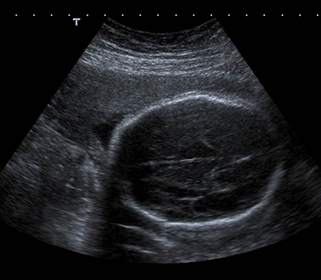
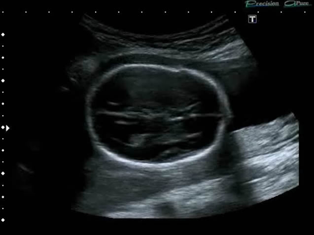
The anatomical landmarks allow the proper positioning of the probe to achieve the optimum section for BPD measurement.
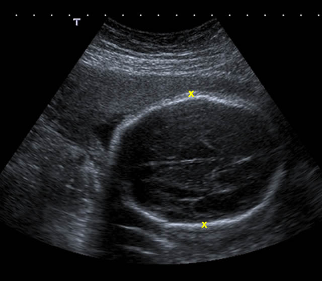
The measurement is made from the outer wall of the skull on the side nearest to the probe to the inner wall of the skull on the side of the head furthest away from the probe.
Click play to watch the video.
This video shows how the measurement end-points are placed from 'leading edge' (the outside of the fetal skull closest to the probe) to 'leading edge' (the inside of the skull furthest from the probe). This is a standardised approach to the measurement of the fetal head and must be performed in all cases to ensure that the measurements can be reproduced throughout the pregnancy. All BPD measurement charts have been developed using this method.
Note: Normally, the image would be frozen for the BPD measurement to be made but we have left the video running to illustrate the positioning of the measurement end points.